Autism children brain imaging is revealing fascinating insights into the neurological underpinnings of autism spectrum disorder (ASD). Researchers are using various brain imaging techniques to explore the unique brain structures and functions in autistic children, comparing them to neurotypical counterparts. This exploration delves into the intricacies of brain development, highlighting structural and functional differences, and shedding light on the potential impact of early intervention and the complex interplay of genetics and environment.
This journey examines the different brain imaging techniques employed in autism research, from structural MRI to functional MRI and diffusion tensor imaging (DTI). We’ll analyze their strengths, limitations, and technical aspects, while acknowledging the ethical considerations involved in studying young brains. The discussion also considers how these findings can inform clinical practice and pave the way for future research directions.
Introduction to Autism and Brain Imaging
Autism spectrum disorder (ASD) is a complex neurodevelopmental condition characterized by persistent challenges in social communication and interaction, as well as restricted, repetitive patterns of behavior, interests, or activities. These characteristics manifest differently in individuals, leading to a wide spectrum of presentations and functional levels. The core features of ASD often emerge in early childhood, though diagnosis can sometimes be delayed.Understanding the neurological underpinnings of ASD is crucial for developing effective interventions and therapies.
Brain imaging techniques offer a window into the brain’s structure and function, providing valuable insights into potential neurological differences in individuals with ASD. This exploration examines the use of brain imaging in autism research, highlighting both the progress made and the ongoing challenges.
Different Brain Imaging Techniques
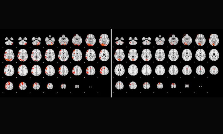
Various neuroimaging techniques are employed to study the brain in individuals with ASD. Structural neuroimaging, such as magnetic resonance imaging (MRI), provides detailed images of brain anatomy. Functional neuroimaging, including functional MRI (fMRI) and electroencephalography (EEG), examines brain activity and function. Diffusion tensor imaging (DTI) assesses the integrity of white matter pathways, crucial for communication between different brain regions.
Historical Context of Brain Imaging in Autism Research
Early studies using brain imaging in autism research focused primarily on identifying structural differences in the brain. As technology advanced, functional imaging techniques allowed researchers to examine brain activity during specific tasks, providing insights into the functional neural correlates of ASD. These advancements have led to a deeper understanding of potential neural pathways and mechanisms implicated in the disorder.
Early studies, though sometimes limited by sample size and methodological constraints, laid the groundwork for more sophisticated and comprehensive investigations.
Limitations and Ethical Considerations
Brain imaging studies involving children present unique challenges. The developing brain undergoes significant structural and functional changes, which can confound the interpretation of observed differences. Furthermore, obtaining informed consent from young participants requires careful consideration of their capacity to understand the study procedures. Ethical guidelines regarding data privacy and confidentiality must also be meticulously followed. The sensitivity and invasiveness of some techniques also necessitate careful consideration and justification.
These factors necessitate stringent methodological rigor and ethical oversight.
Role of Brain Imaging in Understanding the Neurological Basis of Autism
Brain imaging studies have revealed potential neural differences in individuals with ASD, though the exact nature of these differences and their relationship to the core features of the disorder remain areas of ongoing research. Studies have shown potential differences in brain size and structure, such as alterations in certain cortical areas. Functional imaging has also identified variations in brain activity during social interaction and other tasks relevant to autism.
By pinpointing these differences, brain imaging contributes significantly to understanding the neurological basis of autism. This knowledge can, in turn, guide the development of more targeted interventions and treatments.
Brain Imaging Techniques for Autism in Children
Understanding the intricacies of autism spectrum disorder (ASD) in children relies heavily on advanced brain imaging techniques. These methods provide invaluable insights into the structural and functional differences in the brains of children with ASD, offering a window into potential causes and mechanisms underlying the disorder. By comparing these images with those of neurotypical children, researchers can identify patterns and anomalies that might be associated with the unique challenges faced by children with ASD.
Brain imaging studies on autistic children are fascinating, but what if we could get a clearer picture of their brain function using something like a wearable sensor that measures glucose and alcohol levels? A wearable sensor that measures glucose alcohol could it become a reality ? Such a device might offer valuable insights, potentially helping us understand the complex neurochemical processes at play in autism.
Ultimately, these advancements could lead to more targeted and effective interventions for autistic children.
Structural MRI
Structural Magnetic Resonance Imaging (sMRI) is a cornerstone technique in studying the brain. It captures detailed images of the brain’s anatomy, revealing the size, shape, and volume of various brain regions. In the context of autism research, sMRI helps identify potential differences in brain structures, such as the size of the amygdala, hippocampus, or prefrontal cortex, between autistic and neurotypical children.
This technique allows researchers to examine the intricate folds and convolutions of the cerebral cortex, potentially identifying atypical patterns that correlate with ASD characteristics.
- Data acquisition in sMRI involves placing the child within a strong magnetic field. Radio waves excite hydrogen atoms in the brain, and the resulting signals are processed to generate detailed images. Specialized sequences are used to optimize the quality of anatomical information. Careful consideration is given to minimizing motion artifacts, which can arise from patient movement during the scan.
- Preprocessing of sMRI data is crucial for accurate analysis. This often involves correcting for head motion, normalizing brain size to account for individual differences, and segmenting different brain tissues (gray matter, white matter, cerebrospinal fluid). Sophisticated algorithms are employed to minimize the impact of noise and artifacts.
- Analysis of sMRI data focuses on quantifying differences in brain volume or cortical thickness between groups. Statistical methods are used to determine if observed differences are statistically significant, meaning unlikely to be due to chance. Advanced techniques like voxel-based morphometry (VBM) are employed to identify specific regions exhibiting significant differences.
Functional MRI
Functional Magnetic Resonance Imaging (fMRI) measures brain activity by detecting changes in blood flow. Increased neural activity in a particular brain region leads to increased blood flow to that region. fMRI provides insights into the functional connectivity and activity patterns in the brains of autistic children during various tasks or conditions. This is crucial for understanding how different brain regions interact and coordinate to produce behavior.
- Data acquisition in fMRI typically involves the use of blood oxygen level-dependent (BOLD) contrast. The changes in blood oxygenation are measured as the brain performs a specific task or is presented with a stimulus. Multiple scans are acquired over time, capturing the dynamic changes in brain activity. Careful control of experimental conditions is vital to ensure accurate and reliable results.
Brain imaging studies on autistic children are fascinating, but it’s important to remember that there’s still a lot we don’t understand. Recent research, however, is shedding light on the complex neural pathways involved. With the recent announcement that if you received the Pfizer or Moderna vaccines, expect to get a booster starting this fall here , it’s clear that vaccine research and development are ongoing.
Ultimately, continued research into the neurological aspects of autism is crucial for better understanding and care.
- Preprocessing of fMRI data includes motion correction, slice timing correction, spatial normalization, and smoothing. These steps aim to minimize artifacts and ensure that brain activity is analyzed across the entire group in a consistent manner. This is a complex process and often requires specialized software.
- Analysis of fMRI data often involves identifying regions with correlated activity patterns. Researchers look for differences in activation patterns between autistic and neurotypical children during specific tasks, like social interactions or cognitive processing. Statistical parametric mapping (SPM) and other techniques are employed to pinpoint significant differences in brain activity.
Diffusion Tensor Imaging (DTI)
Diffusion Tensor Imaging (DTI) is used to visualize white matter tracts in the brain. White matter tracts are bundles of axons that connect different brain regions, enabling communication between them. DTI allows researchers to study the integrity and organization of these pathways in children with ASD.
- DTI data acquisition relies on measuring the diffusion of water molecules along white matter tracts. The direction and degree of diffusion are used to create images representing the white matter pathways. Advanced imaging techniques like qDTI (quantitative DTI) are employed to study more specific aspects of white matter microstructure.
- Preprocessing of DTI data involves similar steps as sMRI and fMRI, such as motion correction, normalization, and smoothing. Specific preprocessing steps tailored to DTI data are also required to handle unique challenges in this technique. This often involves correcting for distortions and noise that can affect the accuracy of diffusion measurements.
- Analysis of DTI data often involves measuring the fractional anisotropy (FA) and other metrics to assess the integrity of white matter tracts. Researchers may look for differences in the connectivity strength or organization of white matter pathways between autistic and neurotypical children. Graph theoretical analysis can be employed to understand the global brain network structure.
Potential Biases in Brain Imaging Studies
It’s crucial to acknowledge potential biases in brain imaging studies of ASD. Factors like sample size, participant characteristics, and methodological differences can influence the findings. For instance, differences in socioeconomic status, comorbidities, and the specific diagnostic criteria used can all contribute to potential bias.
Summary Table
| Technique | Resolution | Application | Limitations |
|---|---|---|---|
| Structural MRI | High anatomical detail | Brain structure analysis, volume comparisons, cortical thickness | Limited in capturing dynamic processes, susceptible to motion artifacts |
| Functional MRI | Temporal resolution (seconds) | Brain activity during tasks, functional connectivity | Lower spatial resolution compared to sMRI, indirect measure of neural activity |
| Diffusion Tensor Imaging (DTI) | High resolution of white matter tracts | White matter integrity, connectivity analysis | Susceptible to noise and artifacts, limited in measuring gray matter |
Brain Structure and Function Differences

Autism Spectrum Disorder (ASD) is characterized by a range of social, communication, and behavioral challenges. While the exact causes of ASD remain elusive, research strongly suggests that differences in brain structure and function play a crucial role. Understanding these differences is essential for developing effective interventions and therapies.Neuroimaging techniques, such as MRI and fMRI, provide invaluable insights into the brains of individuals with ASD, revealing patterns that distinguish them from neurotypical individuals.
These differences are not necessarily indicators of dysfunction, but rather variations in the way the brain is organized and operates.
Structural Differences in the Brain
Studies have consistently shown structural variations in the brains of children with ASD compared to neurotypical children. These differences often involve the size and shape of various brain regions, potentially impacting the efficiency of information processing and communication within the brain.
- Amygdala: The amygdala is a brain region implicated in processing emotions, particularly fear and anxiety. Research indicates that the amygdala may be larger or show different activity patterns in children with ASD, potentially contributing to heightened sensitivity to sensory stimuli or social cues.
- Hippocampus: The hippocampus is crucial for learning and memory. Studies suggest variations in hippocampal size and activity in children with ASD, potentially impacting their ability to form and retrieve memories, particularly those related to social interactions.
- Prefrontal Cortex: The prefrontal cortex is involved in higher-level cognitive functions such as planning, decision-making, and executive function. Research indicates potential differences in the structure and function of the prefrontal cortex in children with ASD, which could contribute to difficulties with social interaction and flexibility in thinking.
Functional Differences in Brain Activity
Beyond structural differences, functional differences in brain activity are also observed. These differences often relate to specific cognitive functions that are affected in ASD.
- Social Cognition: Children with ASD often exhibit difficulties in understanding social cues and interpreting others’ intentions. Neuroimaging studies show altered activity in brain regions associated with social cognition, such as the superior temporal sulcus (STS), which plays a role in recognizing and interpreting facial expressions and body language.
- Communication: Difficulties with communication are a hallmark of ASD. Neuroimaging studies reveal variations in brain activity in regions involved in language processing, such as Broca’s and Wernicke’s areas, potentially impacting language development and comprehension.
- Sensory Processing: Sensory processing differences are frequently reported in individuals with ASD. Studies show altered activity in brain regions involved in sensory integration, such as the thalamus and somatosensory cortex, potentially contributing to heightened or diminished responses to sensory stimuli.
Developmental Trajectories of Brain Regions
The developmental trajectories of brain regions in children with ASD can differ from those in neurotypical children. These differences might manifest in variations in the timing of brain maturation and the rate of synaptic pruning.
Specific Brain Regions and Associated Behavioral Traits
| Brain Region | Potential Functional Differences | Behavioral Implications |
|---|---|---|
| Amygdala | Potentially larger or showing heightened activity in response to social stimuli, leading to increased anxiety or difficulty with social situations. | Potential for heightened sensitivity to social cues, leading to social withdrawal, difficulty with social interactions, or heightened emotional reactivity. |
| Hippocampus | Variations in size and activity related to memory processing, potentially affecting the ability to learn and retain information, particularly in social contexts. | Difficulties with memory, including social memory, learning new social skills, or remembering social rules and expectations. |
| Prefrontal Cortex | Variations in structure and activity related to executive functions, potentially impacting planning, organization, and problem-solving. | Challenges with planning, organization, and flexibility in thinking, leading to difficulties with tasks requiring these skills. |
Neurodevelopmental Aspects: Autism Children Brain Imaging
Brain imaging studies are offering unprecedented insights into the neurodevelopmental processes underlying autism spectrum disorder (ASD). By observing the structure and function of the developing brain, researchers can identify patterns and potential causes for the unique characteristics of ASD. These findings are crucial for understanding the trajectory of brain development in children with ASD, allowing for a more nuanced approach to early intervention and personalized care.The emerging field of neurodevelopmental research in ASD is shedding light on how the brain develops differently in autistic children.
This knowledge allows us to examine the interplay of genetic predispositions and environmental factors in shaping these differences. Moreover, the ability to track brain development through various stages provides critical information on the effectiveness of early interventions and how they can influence brain structure and function.
How Brain Imaging Illuminates Neurodevelopmental Processes, Autism children brain imaging
Brain imaging techniques, such as fMRI and structural MRI, provide detailed visualizations of the brain’s structure and function. These techniques allow researchers to observe changes in brain volume, connectivity, and activity over time. This longitudinal approach provides crucial data on the developmental trajectory of the brain in children with ASD. Researchers can compare the neurodevelopmental patterns in children with ASD to those in neurotypical children, identifying potential developmental milestones that are reached differently or at varying speeds.
Brain imaging studies on autistic children are fascinating, revealing unique neural pathways and patterns. It’s interesting to reflect on how, just like understanding the intricacies of the autistic brain, it took a pandemic for me to finally admit I have anxiety – why that’s a good thing here. Ultimately, both highlight the importance of self-awareness and acceptance in navigating complex human experiences, and the power of understanding these differences in the broader context of human development.
Impact of Early Intervention on Brain Development
Early intervention programs for children with ASD can have a profound impact on brain development. Studies suggest that early intervention can lead to improvements in social-emotional skills, communication, and cognitive abilities. These interventions can potentially influence brain structure and function by stimulating neural pathways and promoting adaptive behaviors. The effects of early intervention on brain development are a critical area of ongoing research, with the goal of optimizing outcomes for children with ASD.
Role of Genetics and Environment in Shaping Brain Structure and Function
The development of the brain in children with ASD is a complex interplay of genetic and environmental factors. Genetic predisposition plays a significant role in increasing the risk of developing ASD. This genetic influence is evidenced by the higher prevalence of ASD in families with a history of the disorder. However, environmental factors, including prenatal exposures and early life experiences, also contribute to shaping brain structure and function.
Interplay Between Genetics and Environmental Factors
The interplay between genetics and environmental factors is critical to understanding the development of ASD. Genetic factors may create a predisposition to certain brain characteristics, but environmental influences can modulate how these genetic predispositions manifest. For example, a child with a genetic predisposition for difficulties with social interaction might exhibit more pronounced symptoms if exposed to stressful environments or lack supportive social interactions.
Methods to Assess Environmental Factors’ Impact
Several methods are used to assess the impact of environmental factors on brain development in children with ASD. These methods often combine brain imaging techniques with questionnaires and interviews to gather data on the child’s experiences. For instance, researchers may assess the impact of prenatal exposures, such as maternal stress or exposure to toxins, on brain structure and function using MRI.
Other methods involve tracking early childhood experiences and environmental factors to determine potential correlations with brain development in children with ASD. The goal is to identify specific environmental factors that may exacerbate or mitigate the impact of genetic predispositions on brain development.
Clinical Implications and Future Directions
Brain imaging studies are offering a window into the complexities of autism spectrum disorder (ASD) in children, revealing potential biomarkers and neural correlates that could revolutionize clinical practice. These insights are moving beyond simple observation to offer more targeted interventions and personalized approaches. The quest to understand the intricacies of autism’s neurobiology is not just academic; it holds the key to improving the lives of children with ASD and their families.Understanding the brain’s role in ASD is crucial for developing better strategies for diagnosis, treatment, and support.
This understanding is not merely theoretical; it has the potential to translate into concrete improvements in the lives of children affected by ASD. By examining brain structure and function, we can potentially uncover early indicators, tailor interventions to individual needs, and even monitor progress over time.
Clinical Application of Brain Imaging Findings
Brain imaging techniques are increasingly used to identify subtle differences in brain structure and function that may correlate with ASD. These differences, while not always definitive, can provide valuable insights for clinicians. For example, studies have shown altered connectivity patterns in specific brain regions, which might be indicative of social communication difficulties. This knowledge can help clinicians identify children at risk and provide targeted interventions to address potential deficits early on.
Diagnostic Criteria Based on Brain Imaging Data
While current diagnostic criteria for ASD rely primarily on behavioral observations, brain imaging data could potentially augment or refine these criteria. This is an active area of research. Future diagnostic tools might incorporate neuroimaging findings to create a more comprehensive assessment, potentially leading to earlier and more accurate diagnoses. This could include evaluating specific brain regions and neural pathways implicated in social cognition, communication, and repetitive behaviors.
Monitoring Treatment Response Using Brain Imaging
Brain imaging has the potential to monitor the effectiveness of various treatments for ASD. For instance, by tracking changes in brain activity or connectivity patterns following therapy, clinicians could determine if an intervention is having a positive impact. This dynamic approach allows for adjustments to treatment plans based on real-time brain responses, fostering personalized care and potentially optimizing outcomes.
Open Questions in Autism Brain Imaging Research
The field of autism brain imaging is continually evolving, and several critical questions remain unanswered. These questions encompass the need for standardized protocols, the reproducibility of findings across different studies, and the long-term implications of these neuroimaging findings.
- What are the specific neural mechanisms underlying the diverse symptoms of autism?
- How do environmental factors and genetic predispositions interact to influence brain development in individuals with ASD?
- Can we identify specific brain imaging biomarkers for ASD that are reliable and sensitive enough for widespread clinical use?
- How can we best integrate brain imaging data into existing diagnostic and treatment approaches?
Future Research Directions
Future research in autism brain imaging should focus on developing more sophisticated methodologies to study brain function and connectivity. This involves leveraging advanced techniques like diffusion tensor imaging (DTI) to study white matter pathways, or functional magnetic resonance imaging (fMRI) to examine brain activity in response to social stimuli.
- Longitudinal studies are crucial to understand the progression of brain development and how it relates to the emergence and evolution of autism symptoms over time.
- Research should also explore the use of machine learning algorithms to analyze large datasets of brain imaging data, potentially leading to the development of more sophisticated diagnostic tools and individualized treatment strategies.
- Cross-sectional studies are important to compare brain imaging features between different age groups and subtypes of autism.
- Collaboration between researchers, clinicians, and families is essential to ensure that research findings translate into practical applications and improved support for individuals with ASD.
Outcome Summary

In conclusion, autism children brain imaging offers a compelling window into the complex neurological landscape of ASD. The research reveals significant structural and functional differences in the brains of autistic children, highlighting the intricate interplay of genetics, environment, and neurodevelopment. This knowledge is crucial for understanding the condition and for developing more effective interventions and therapies. Further research, guided by ethical considerations, promises to uncover even more about the intricacies of the autistic brain, leading to improved diagnostics and treatment strategies for children with ASD.