Superficial muscles of neck – Superficial muscles of the neck are a fascinating group of muscles that play a crucial role in head and neck movement, posture, and even facial expressions. They’re situated just beneath the skin, giving them a superficial location, and work in concert with other structures in the neck to perform a wide range of actions. Understanding their individual functions, interactions, and clinical significance is essential for comprehending the complex mechanics of the neck.
This exploration delves into the anatomy, function, and clinical implications of these vital muscles. We’ll examine their origins, insertions, actions, and how they work together to produce specific movements. From the sternocleidomastoid’s role in head turning to the trapezius’s contribution to posture, we’ll uncover the intricate workings of the superficial neck muscles. Tables will visually summarize key anatomical data, and illustrative examples will further enhance understanding.
Introduction to Superficial Muscles of the Neck
The superficial muscles of the neck are a group of muscles located just beneath the skin and superficial to deeper layers of neck musculature. These muscles play a crucial role in various movements and functions of the head and neck, including posture, facial expression, and swallowing. Understanding their anatomical relationships and functions is essential for comprehending the overall mechanics of the neck.
Ever wondered about those muscles in your neck? The superficial ones, like the sternocleidomastoid, are crucial for head movement and posture. Recent research suggests that subtle changes in the brain, like those potentially linked to Alzheimer’s disease, might be detectable as early as 30 years before the first symptoms appear. can alzheimers be detected 30 years before it appears This could lead to earlier intervention, which is fascinating and might have implications for strengthening these neck muscles as a preventative measure.
Ultimately, understanding the complex interplay between physical health and neurological well-being is crucial for proactive health choices.
Their superficial location makes them readily palpable, allowing for assessment of their condition in clinical settings.These muscles are interconnected and often work synergistically to produce coordinated movements. Their origins and insertions, along with the surrounding structures, determine their specific actions. Knowing the actions of these muscles provides insight into potential impairments or dysfunction, aiding in diagnosis and treatment.
Anatomical Location and Relationships
The superficial muscles of the neck are situated in the anterior, lateral, and posterior regions of the neck. They lie superficial to deeper structures like the platysma, sternocleidomastoid, and trapezius. Their location allows for relatively easy access and palpation, which is valuable in physical examinations. The relationships with deeper muscles, blood vessels, and nerves contribute to the intricate mechanical system of the neck.
Comparative Analysis of Major Superficial Neck Muscles
A clear understanding of the anatomical details of each muscle is essential for comprehending their specific functions. The table below summarizes the location, origin, insertion, and actions of the key superficial neck muscles.
| Muscle | Location | Origin | Insertion | Action |
|---|---|---|---|---|
| Platysma | Anterior neck, superficial to other neck muscles | Fascia over pectoralis major and deltoid muscles | Mandible, lower face, and skin of the neck | Draws down the lower lip and angles of the mouth, tenses skin of the neck, and assists in depressing the mandible |
| Sternocleidomastoid | Anterior lateral neck | Manubrium of sternum and medial third of clavicle | Mastoid process of temporal bone and superior nuchal line of occipital bone | Flexes the neck laterally, rotates the head to the opposite side, and elevates the sternum during forced inhalation. |
| Trapezius | Posterior neck and upper back | External occipital protuberance, superior nuchal line, ligamentum nuchae, and spinous processes of C7-T12 vertebrae | Lateral third of clavicle, acromion, and spine of scapula | Elevates, retracts, and rotates the scapula, and extends the head and neck. |
Individual Muscle Descriptions
Diving deeper into the anatomy of the neck, we’ll now explore the individual muscles that make up its superficial layer. Understanding their origins, insertions, innervations, and functions is crucial for comprehending the complex mechanics of head and neck movement. From the powerful sternocleidomastoid to the subtle platysma, each muscle plays a specific role in posture, movement, and even facial expression.
Sternocleidomastoid Muscle
The sternocleidomastoid, a prominent muscle on either side of the neck, is a key player in head and neck movements. Its origin spans across the sternum and clavicle, specifically the manubrium of the sternum and medial third of the clavicle. The insertion point is located on the mastoid process of the temporal bone and superior nuchal line of the occipital bone.
Ever wondered about the superficial muscles of the neck? They’re crucial for head and neck movement, but their function isn’t the only thing to consider. Just like how not all dietary fibers are equal heres why , different muscle types respond better to certain nutrients. Understanding how nutrition supports muscle health is a vital part of overall neck health and well-being.
Innervation comes from the accessory nerve (cranial nerve XI) and the cervical plexus. Crucially, this muscle is responsible for flexion of the neck, rotation of the head to the opposite side, and lateral flexion of the neck. For example, when you tilt your head to one side, the sternocleidomastoid on that side contracts, pulling the head in the desired direction.
Trapezius Muscle
The trapezius, a broad, superficial muscle spanning the upper back and neck, plays a significant role in posture and movement of the shoulder girdle. Originating from the occipital bone, the ligamentum nuchae, and the spinous processes of the thoracic vertebrae, its fibers converge to insert on the lateral third of the clavicle, acromion process, and spine of the scapula.
Innervation is provided by the accessory nerve (cranial nerve XI) and upper cervical nerves. The trapezius facilitates scapular elevation, depression, retraction, and rotation, and is essential for maintaining proper posture and allowing for a wide range of shoulder movements. A weakened trapezius can lead to postural imbalances and shoulder pain.
Platysma Muscle
The platysma, a thin, sheet-like muscle extending from the lower face to the neck, plays a subtle yet important role in facial expressions and neck movements. Its origin is located in the fascia covering the pectoralis major and deltoid muscles. It inserts into the mandible and skin of the lower face, including the mouth. The platysma is innervated by the cervical branch of the facial nerve (cranial nerve VII).
Its action is primarily responsible for depressing the mandible, lowering the lower lip, and contributing to facial expressions like frowning or pursing the lips.
Subclavius Muscle
The subclavius, a small muscle situated beneath the clavicle, plays a critical role in stabilizing the shoulder. Originating from the first rib, it inserts on the inferior surface of the clavicle. It’s innervated by the nerve to the subclavius, a branch of the upper trunk of the brachial plexus. The primary action of the subclavius is to depress the clavicle, and assist in stabilizing the shoulder joint during movements.
This is crucial for maintaining the position of the shoulder girdle.
Omohyoid Muscle
The omohyoid muscle, a two-bellied muscle located in the neck, assists in lowering the hyoid bone. Its origin is on the superior border of the scapula, and its insertion is on the body of the hyoid bone. The omohyoid is innervated by the cervical plexus. Its primary action is to depress the hyoid bone, which is important for swallowing and other neck movements.
Scalene Muscles
The scalene muscles (anterior, middle, and posterior) are three important muscles located in the neck that contribute to respiration and neck stability.
| Muscle | Origin | Insertion | Innervation | Action |
|---|---|---|---|---|
| Anterior Scalene | Transverse processes of cervical vertebrae (C3-C6) | First rib | Cervical plexus (C3-C8) | Elevates first rib, flexes and laterally flexes neck |
| Middle Scalene | Transverse processes of cervical vertebrae (C2-C7) | First rib | Cervical plexus (C3-C8) | Elevates first rib, flexes and laterally flexes neck |
| Posterior Scalene | Transverse processes of cervical vertebrae (C5-C7) | Second rib | Cervical plexus (C3-C8) | Elevates second rib, flexes and laterally flexes neck |
Muscle Interactions and Synergies: Superficial Muscles Of Neck
The superficial muscles of the neck are not isolated units; they work in concert to produce coordinated movements of the head and neck. Understanding their interactions, both synergistic and antagonistic, is crucial for comprehending how these muscles contribute to posture and movement. This interplay allows for smooth and controlled actions, preventing unwanted or excessive movement.
Sternocleidomastoid and Trapezius Action in Head Movements
The sternocleidomastoid and trapezius muscles are key players in head movements. The sternocleidomastoid, acting bilaterally, flexes the neck and head forward. Unilaterally, it rotates the head towards the opposite side and tilts it laterally. The trapezius, on the other hand, plays a multifaceted role. It extends the head and neck, and when acting unilaterally, rotates the head towards the same side and tilts it laterally.
These contrasting actions, when combined, create a wide range of controlled head movements.
Synergistic and Antagonistic Relationships
The superficial neck muscles exhibit both synergistic and antagonistic relationships. Synergistic muscles work together to achieve a particular movement. For instance, during flexion of the neck, the sternocleidomastoids of both sides act synergistically. Antagonistic muscles oppose each other’s actions, allowing for controlled movement. The sternocleidomastoid and splenius capitis are antagonists in head flexion, as the splenius capitis extends the head.
This interplay of synergistic and antagonistic actions is essential for smooth and controlled movement.
Role in Maintaining Posture
The superficial neck muscles are vital for maintaining an upright posture. By constantly adjusting tension and working in coordination, these muscles counteract the forces of gravity, ensuring the head and neck remain stable. The interplay of the trapezius, sternocleidomastoid, and other superficial neck muscles is essential for supporting the head and maintaining an erect posture, which is critical for overall body balance and stability.
Muscle Interactions Table, Superficial muscles of neck
| Muscle Pair | Synergy | Antagonism | Movement |
|---|---|---|---|
| Sternocleidomastoid (bilateral) | Sternocleidomastoid (bilateral) | Splenius capitis, Semispinalis capitis | Neck flexion |
| Sternocleidomastoid (unilateral) | Scalenes | Sternocleidomastoid (contralateral), Splenius capitis | Head rotation and lateral flexion (to the same side) |
| Trapezius (upper fibers) | Sternocleidomastoid | Longus capitis, Longus colli | Head extension |
| Trapezius (middle fibers) | Splenius capitis | Sternocleidomastoid | Scapular elevation and retraction |
| Trapezius (lower fibers) | Rhomboids | Levator scapulae | Scapular depression and downward rotation |
Clinical Significance
The superficial muscles of the neck, though seemingly small, play a crucial role in maintaining head posture, facilitating movement, and contributing to overall neck health. Understanding their clinical significance is vital for diagnosing and treating various conditions affecting the neck region. Disruptions to these muscles can manifest in a variety of ways, impacting not only movement but also potentially leading to pain and discomfort.Muscle weakness or injury to the superficial neck muscles can significantly affect posture and movement.
For instance, a weakened sternocleidomastoid muscle could lead to an altered head position, potentially causing strain on the cervical spine. Conversely, a strained or damaged muscle, such as the trapezius, could result in localized pain and restricted range of motion.
Muscle Imbalances and Their Implications
Muscle imbalances in the neck, often resulting from repetitive movements, poor posture, or underlying medical conditions, are a common cause of neck pain. These imbalances can occur when certain muscles become overly tight and strong while others weaken, leading to a compensatory mechanism that further stresses the affected area. For example, prolonged computer work often leads to tight, overactive upper trapezius muscles, while the deeper neck muscles may weaken, creating an imbalance.
This imbalance can contribute to a forward head posture, putting increased strain on the cervical spine.
Potential Causes of Muscle Imbalances
Several factors contribute to muscle imbalances in the neck region. Repetitive stress, such as prolonged periods of sitting or working at a computer, can lead to overactivity of specific muscles. Poor posture, characterized by slouching or a forward head posture, can strain certain neck muscles and weaken others. Underlying medical conditions, such as arthritis or neurological disorders, can also influence muscle strength and function.
Stress and anxiety can also contribute to muscle tension and tightness in the neck, impacting muscle balance. Furthermore, inadequate sleep, poor nutrition, or insufficient physical activity can impact muscle health and potentially contribute to imbalances.
Ever wondered about the superficial muscles of the neck? They’re crucial for head movement and posture, playing a vital role in daily life. Interestingly, some research suggests that understanding how our bodies react to certain illnesses, like how immunotherapy works against melanoma, could potentially lead to better treatment strategies for muscle-related issues. For example, exploring how does immunotherapy work melanoma might uncover broader insights into the body’s immune response, which could then be applied to the study of the superficial muscles of the neck.
Ultimately, these intricate connections between different areas of health are fascinating to explore.
Contribution to Headaches and Neck Pain
Muscle imbalances and tightness in the superficial neck muscles are frequently associated with headaches and various neck pain conditions. The strain and tension placed on the surrounding tissues and structures can trigger pain signals, leading to headaches. Furthermore, the restricted movement resulting from muscle imbalances can impede proper blood flow and nutrient delivery to the muscles and nerves, exacerbating pain and discomfort.
For instance, a tight trapezius muscle can refer pain to the head, potentially manifesting as a tension headache. Similarly, a weakened deep neck flexor muscle can contribute to a forward head posture, causing persistent neck pain.
Common Clinical Conditions
- Cervical Spondylosis: This degenerative condition of the cervical spine can result in stiffness, pain, and reduced mobility in the neck. It can also be associated with muscle strain and imbalances.
- Whiplash: A sudden, forceful impact on the neck can cause whiplash, leading to injuries in the muscles, ligaments, and tendons of the neck. Muscle strain and imbalances are common consequences.
- Tension Headaches: These headaches, often accompanied by neck pain, are frequently linked to muscle tension and imbalances in the superficial neck muscles.
- Myofascial Pain Syndrome: This condition involves trigger points in the muscles, which can lead to pain radiating to other areas of the body, including the head and neck. Tightness and imbalances in superficial neck muscles are often a contributing factor.
Illustrative Examples
Diving deeper into the superficial neck muscles reveals a fascinating interplay of form and function. These muscles, often overlooked, play crucial roles in head and neck movement, posture, and even breathing. Understanding their locations, relationships, and interactions provides a clearer picture of their significance in overall health.
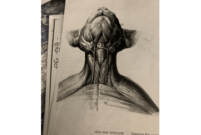
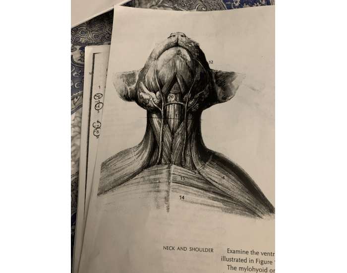
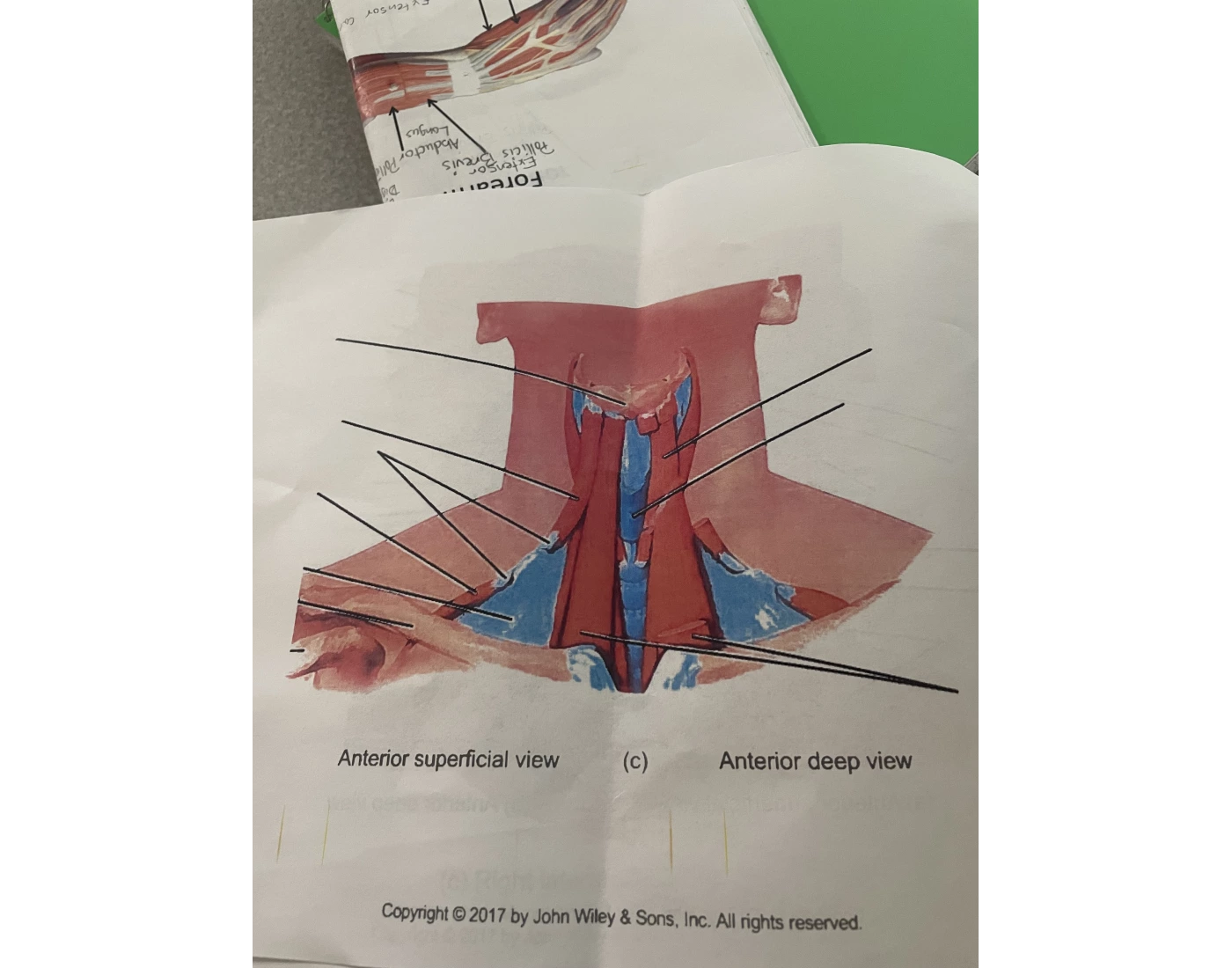
Detailed Illustration of Superficial Neck Muscles
The superficial neck muscles form a complex network, with several key players contributing to the overall structure and function. The sternocleidomastoid, a prominent muscle, originates from the sternum and clavicle, extending diagonally upward to insert on the mastoid process of the temporal bone. The trapezius, another substantial muscle, spans the posterior neck, extending from the occipital bone and vertebrae down to the scapula.
Other muscles, like the platysma, lie more superficially, contributing to the overall aesthetic of the neck. These muscles interweave, their fibers oriented in different directions, creating a tapestry of support and movement. Imagine a layered structure, with deeper muscles providing stability, while the superficial muscles contribute to dynamic movements.
Movement During Specific Actions
The interplay of superficial neck muscles becomes evident during various movements. For instance, turning the head relies on the coordinated action of the sternocleidomastoid muscles on both sides. Contraction of one side rotates the head in the opposite direction. Shrugging the shoulders involves the trapezius muscle, its superior fibers elevating the scapula. The platysma, while not directly involved in these movements, contributes to the overall tension and appearance of the neck during these actions.
Understanding the precise actions of each muscle allows for a more comprehensive understanding of how the neck functions.
Healthy vs. Imbalanced Neck
Visualizing a healthy neck reveals a symmetrical structure, with muscles appearing balanced and toned. The sternocleidomastoid muscles on both sides exhibit similar development, and the trapezius muscle displays even strength across its various sections. Conversely, an imbalanced neck may exhibit asymmetry in muscle development, often associated with postural deviations or repetitive strain injuries. One sternocleidomastoid muscle might be more prominent or tense than the other, or the trapezius might show signs of overuse on one side, potentially leading to head tilt or shoulder asymmetry.
The illustration of these contrasting scenarios highlights the importance of maintaining muscular balance in the neck region.
Role in Respiration
While not the primary respiratory muscles, the superficial neck muscles indirectly participate in respiration. The sternocleidomastoid muscles, during forced inhalation, assist in elevating the sternum and rib cage, increasing the volume of the thoracic cavity. The trapezius, although less directly involved, still contributes to the overall stability of the upper torso, which indirectly affects respiratory mechanics. During deep breaths or strenuous activities, these muscles may contribute more significantly.
Understanding this indirect role adds to the overall comprehension of the neck’s functions.
Muscle Fiber Orientation
The superficial neck muscles exhibit a diverse array of fiber orientations. The sternocleidomastoid’s fibers run diagonally, enabling its rotational action. The trapezius’s fibers, running in multiple directions, allow for varied movements like elevation, depression, and retraction of the scapula. The platysma’s fibers run horizontally, contributing to its role in facial expressions and neck movements. These varying orientations, akin to a complex web, enable the precise and varied movements of the neck and head.
The visual representation of these fiber patterns highlights the complexity and efficiency of the neck’s muscle system.
Last Recap
In conclusion, the superficial muscles of the neck are a critical component of the human body’s intricate system of movement and support. Their diverse functions, from head movement to posture maintenance, highlight their importance in everyday activities. Understanding their interactions, clinical significance, and potential for injury is essential for maintaining optimal neck health. This exploration provides a foundational understanding of these muscles, setting the stage for further investigation into more specialized areas.