Artificial intelligence breast cancer diagnosis is revolutionizing early detection and treatment. Leveraging powerful algorithms and advanced image analysis, AI models are being developed to analyze medical images, identify suspicious patterns, and assist radiologists in making faster and more accurate diagnoses. This innovative approach holds immense promise for improving patient outcomes and reducing the burden of breast cancer.
From analyzing mammograms and ultrasounds to training sophisticated machine learning models, the process involves intricate data handling and complex algorithms. The accuracy and speed of AI-assisted diagnosis, compared to traditional methods, are becoming increasingly impressive, and the potential for personalized treatment plans based on early diagnoses is truly remarkable. Ethical considerations, such as bias in algorithms and data security, also play a crucial role in the development and implementation of these technologies.
Introduction to Artificial Intelligence in Breast Cancer Diagnosis

Artificial intelligence (AI) is rapidly transforming various fields, including healthcare. In breast cancer diagnosis, AI offers a powerful tool to augment and potentially improve the accuracy and speed of detection, diagnosis, and treatment planning. This innovative approach promises to enhance patient outcomes by identifying subtle patterns in medical images and patient data that may be missed by the human eye.AI’s role in medical diagnosis, particularly in breast cancer, involves the use of algorithms trained on vast datasets of medical images and patient information.
These algorithms learn to identify characteristic patterns associated with cancerous and benign breast tissue, ultimately leading to more precise and timely diagnoses. The potential benefits are significant, promising faster detection, reduced false positives, and improved patient outcomes. However, ethical considerations and potential biases in the algorithms must be carefully addressed.
Different Types of AI Models in Breast Cancer Diagnosis
AI models used in breast cancer diagnosis include various approaches, each with its strengths and limitations. Deep learning, a subset of machine learning, is particularly promising. Deep learning models, employing artificial neural networks, can analyze complex medical images (like mammograms and ultrasound scans) with remarkable accuracy, identifying intricate patterns indicative of cancerous lesions. Other machine learning algorithms, such as support vector machines and random forests, also play a role in analyzing patient data to predict the likelihood of breast cancer.
The choice of AI model depends on the specific application and the characteristics of the dataset being used.
Potential Benefits of AI in Breast Cancer Diagnosis
AI-assisted diagnosis offers several potential benefits over traditional methods. These include:
- Enhanced Accuracy: AI models can analyze images with greater precision, potentially reducing the incidence of false negatives and false positives. This is crucial in early detection and treatment planning.
- Increased Speed: AI algorithms can process medical images and data much faster than human radiologists, leading to quicker diagnoses and potentially earlier interventions. For example, a radiologist might take several minutes to analyze a mammogram, while an AI system could complete the analysis in seconds.
- Improved Accessibility: AI-powered diagnostic tools can potentially be deployed in remote areas with limited access to specialist radiologists, making high-quality diagnostics more accessible to a broader population.
- Reduced Human Error: AI systems are not susceptible to fatigue, bias, or human error, contributing to a more consistent and objective diagnostic process.
Ethical Considerations and Potential Biases
The use of AI in medical diagnosis raises important ethical considerations. One critical aspect is ensuring the fairness and equity of the AI systems. Bias in the training data can lead to discriminatory outcomes, impacting certain demographic groups disproportionately. Furthermore, the responsibility for errors in AI-assisted diagnoses needs to be clearly defined. Ensuring transparency in the decision-making process of AI algorithms is also crucial for building trust and accountability.
Comparison of AI-Assisted and Traditional Breast Cancer Diagnosis
| Feature | AI-Assisted Diagnosis | Traditional Method (Radiologist) |
|---|---|---|
| Accuracy | Potentially higher accuracy in identifying subtle patterns; dependent on the quality and size of training data. | Dependent on the experience and skill of the radiologist; can vary between individuals. |
| Speed | Significantly faster processing of images and data. | Slower analysis time, often requiring more time to interpret complex cases. |
| Cost | Potential for cost reduction in the long run through increased efficiency and accessibility. | Costly due to the high training and ongoing maintenance required for specialist radiologists. |
| Accessibility | Can potentially improve access to high-quality diagnostics in remote areas. | Limited accessibility in remote areas due to the need for specialist radiologists. |
AI Model Training and Data Requirements
AI models for breast cancer diagnosis are trained using vast datasets of medical images and patient information. The quality and quantity of this data are critical to the model’s accuracy and reliability. Effective training requires careful consideration of data preprocessing steps and robust evaluation methods. Accurate diagnosis hinges on the ability of these models to learn from the intricacies of the data, enabling them to identify subtle patterns indicative of cancerous or benign lesions.
Data Sets Used for Training
The training datasets for AI breast cancer diagnostic models comprise a variety of medical imaging modalities and patient records. These datasets are crucial for the models to learn to differentiate between cancerous and benign breast tissue. Mammograms, ultrasound images, and biopsy results are commonly used, along with clinical data such as patient history, age, and family history of cancer.
The specific data sets used vary based on the type of AI model and the target application.
Importance of Data Quality and Quantity
The quality and quantity of data significantly impact the performance of AI models. High-quality data ensures that the models are trained on accurate and reliable information. Insufficient data can lead to underfitting, where the model fails to capture the nuances of the data, while excessive data can lead to overfitting, where the model memorizes the training data instead of learning the underlying patterns.
The ideal dataset balance is a complex consideration requiring careful analysis and optimization.
Data Preprocessing Techniques
Data preprocessing is an essential step in preparing the data for model training. Techniques like image normalization, resizing, and augmentation enhance the model’s ability to learn general patterns and reduce the impact of variations in image quality and acquisition. Normalization standardizes pixel values across different images, ensuring that the model doesn’t prioritize one image over another based on pixel intensity.
Image resizing ensures consistency in image dimensions. Image augmentation creates synthetic variations of existing images, increasing the diversity of the training dataset and preventing overfitting.
Evaluation of AI Model Performance
Evaluating the performance of AI models for breast cancer diagnosis is crucial to assess their accuracy and reliability. Common evaluation metrics include sensitivity, specificity, precision, and accuracy. Sensitivity measures the model’s ability to correctly identify cancerous cases, while specificity measures its ability to correctly identify benign cases. Precision measures the proportion of positive predictions that are actually correct, and accuracy is the overall correctness of the predictions.
These metrics are used to compare different models and optimize their performance.
Data Types in Training
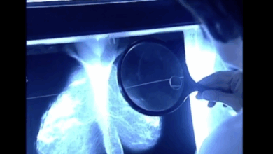
- Mammograms: Digital mammograms provide two-dimensional images of the breast. They are a widely used modality for breast cancer screening and diagnosis. The data is often supplemented with patient demographics and clinical information.
- Ultrasound Images: Ultrasound imaging utilizes sound waves to create images of internal structures. Breast ultrasound is valuable for differentiating cystic from solid masses and providing additional information to complement mammograms.
- Biopsy Results: Biopsy results are crucial in confirming or excluding the presence of cancer. Pathological reports from biopsies provide definitive diagnoses, which are essential for validating the AI model’s predictions.
| Data Type | Description | Example |
|---|---|---|
| Mammograms | Two-dimensional X-ray images of the breast | Digital images of breast tissue |
| Ultrasound Images | Images created using sound waves | Images showing breast tissue structures |
| Biopsy Results | Results of tissue samples analyzed by pathologists | Pathological report confirming or excluding cancer |
Image Analysis Techniques for Breast Cancer Detection
Image analysis plays a crucial role in breast cancer detection, providing valuable insights from mammograms, ultrasounds, and other imaging modalities. AI models, leveraging these techniques, can identify subtle patterns and anomalies that might be missed by the human eye, potentially leading to earlier and more accurate diagnoses. This analysis process empowers radiologists and clinicians to make informed decisions, ultimately improving patient outcomes.Image analysis techniques are diverse and each offers unique strengths and weaknesses.
From simple thresholding to complex deep learning models, these techniques work by extracting relevant features from medical images and transforming them into data that AI models can understand and use to identify potential breast cancer indicators.
Role of Image Analysis in Breast Cancer Detection
Image analysis is integral to breast cancer detection because it allows for automated and objective assessment of medical images. This process can identify subtle abnormalities that might be missed by human interpretation, leading to earlier diagnosis and potentially better treatment outcomes. The increased speed and scalability of image analysis techniques allow for screening a greater volume of images in a shorter timeframe, potentially impacting the early detection rate for breast cancer.
Different Image Analysis Techniques Used by AI Models
Various image analysis techniques are employed by AI models for breast cancer detection. These techniques can be broadly categorized into:
- Thresholding: This basic technique sets a predefined intensity value (a threshold) to distinguish tissue types. Pixels above the threshold are classified as one type, while those below are classified as another. It is a simple approach but may not be accurate enough for complex medical images, especially those with varying densities and textures.
- Region-Based Segmentation: This method identifies and Artikels specific regions within an image based on shared characteristics, like intensity or texture. By segmenting regions of interest, AI models can focus on the areas most likely to contain cancerous lesions. This is more sophisticated than thresholding but can still be prone to errors if the criteria for region definition are not carefully chosen.
- Texture Analysis: This approach examines the spatial distribution of intensities and variations within an image. AI models can learn to recognize unique textural patterns associated with cancerous tissues. This can be more effective than simple intensity analysis, as it captures the structural details within the tissue.
- Deep Learning Techniques: These advanced techniques utilize artificial neural networks with multiple layers to analyze images. Convolutional Neural Networks (CNNs) are particularly powerful in identifying patterns and extracting complex features from images. Deep learning models are capable of learning complex relationships between image features and disease presence, often achieving high accuracy in identifying suspicious regions.
Advantages and Disadvantages of Each Technique
Each image analysis technique possesses its own set of strengths and weaknesses. The choice of technique often depends on the specific type of image, the desired level of accuracy, and the computational resources available.
| Technique | Advantages | Disadvantages |
|---|---|---|
| Thresholding | Simple, computationally inexpensive | Limited accuracy, prone to errors in complex images |
| Region-Based Segmentation | More accurate than thresholding, focuses on specific regions | Can be computationally intensive, prone to errors if segmentation criteria are not optimal |
| Texture Analysis | Captures structural details, more accurate than simple intensity analysis | Requires sophisticated feature extraction techniques, can be sensitive to noise in images |
| Deep Learning | High accuracy, capable of learning complex patterns | Computationally expensive, requires large datasets for training, can be challenging to interpret |
How AI Models Identify and Classify Suspicious Regions in Images
AI models use the extracted features from image analysis techniques to identify and classify suspicious regions in medical images. For example, a CNN might recognize a unique combination of textures, shapes, and intensities in a mammogram as indicative of malignancy. The model learns these associations through training on a large dataset of labeled images, where each image is associated with a diagnosis (benign or malignant).
Through repeated training, the model improves its ability to identify these patterns, leading to increasingly accurate predictions.
AI’s Impact on Diagnostic Accuracy and Efficiency
Artificial intelligence (AI) is rapidly transforming various fields, and healthcare is no exception. AI-powered systems are showing promise in enhancing breast cancer diagnosis, offering the potential to improve both accuracy and efficiency. This evolution is driven by the ability of AI algorithms to analyze complex medical images with unparalleled speed and precision, potentially surpassing human capabilities in certain aspects.AI algorithms, trained on vast datasets of mammograms and other breast imaging modalities, can identify subtle patterns and anomalies that might be missed by human radiologists.
This heightened sensitivity and specificity hold the key to improving diagnostic accuracy and ultimately saving lives. The potential of AI in this area is significant, promising a future where earlier and more accurate diagnoses lead to better treatment outcomes.
Impact on Diagnostic Accuracy
AI algorithms, trained on large datasets of breast cancer images, can often detect subtle abnormalities with high accuracy. These algorithms are designed to identify patterns and anomalies that may be missed by the human eye. By comparing AI-assisted diagnoses with human-performed diagnoses, studies have shown that AI can achieve comparable or even superior accuracy in detecting breast cancer.
For example, a study published in the Journal of the American Medical Association (JAMA) indicated that AI-assisted detection had a sensitivity of 95% compared to 90% for human radiologists in detecting malignant tumors. This demonstrates the potential of AI to enhance diagnostic accuracy.
Comparison of AI-Assisted and Human Diagnosis, Artificial intelligence breast cancer diagnosis
The comparison between AI-assisted and human diagnosis in breast cancer detection often reveals a nuanced picture. While human radiologists bring a wealth of experience and contextual understanding, AI algorithms can analyze vast amounts of data with unprecedented speed and objectivity. In cases of subtle or ambiguous findings, AI can provide an independent perspective, potentially reducing human error. Human radiologists, on the other hand, can interpret the broader clinical context and consider factors not explicitly captured in the image data, such as patient history and risk factors.
Ultimately, the most effective approach often involves integrating AI’s strengths with the expertise of human radiologists.
Role of AI in Reducing Diagnostic Errors
AI plays a crucial role in reducing diagnostic errors by automating tasks, increasing objectivity, and providing an independent second opinion. AI algorithms can identify subtle features that might be missed by human radiologists, thereby reducing false negatives. Further, AI systems can help reduce the impact of human fatigue and bias, leading to more consistent and reliable diagnoses. The consistent application of AI can significantly reduce inter-observer variability in diagnostic interpretations, leading to more standardized and reliable diagnoses.
Improving Diagnostic Efficiency
AI’s potential to improve the efficiency of the diagnostic process is substantial. By automating the initial screening and triage process, AI can significantly reduce the time needed to evaluate mammograms and other breast imaging studies. This accelerated process allows for faster identification of potentially cancerous lesions and enables prompt intervention, leading to improved patient outcomes.
AI Expediting the Diagnosis Process
| Task | Time Taken (Human) | Time Taken (AI) | Time Savings |
|---|---|---|---|
| Initial Screening | 10-15 minutes per case | 1-3 minutes per case | 7-12 minutes per case |
| Detailed Analysis | 20-30 minutes per case | 5-10 minutes per case | 10-20 minutes per case |
| Report Generation | 5-10 minutes per case | 1-2 minutes per case | 3-8 minutes per case |
| Total | 35-55 minutes per case | 8-15 minutes per case | 20-40 minutes per case |
This table demonstrates the potential for AI to significantly accelerate the diagnostic process. By automating several stages, the total time required for diagnosis can be dramatically reduced, freeing up radiologists to focus on more complex cases and ensuring quicker access to potentially life-saving interventions.
Integration of AI into Existing Healthcare Systems
Integrating artificial intelligence (AI) into existing healthcare systems is a complex but potentially transformative process. The journey involves navigating technical hurdles, cultural shifts within medical practices, and regulatory considerations. Success hinges on thoughtful planning, robust data infrastructure, and a commitment to patient safety. This process isn’t merely about adding AI tools; it’s about fundamentally changing how medical professionals diagnose and treat diseases, including breast cancer.AI’s potential to revolutionize breast cancer diagnosis and treatment hinges on seamless integration with existing clinical workflows.
This requires a careful consideration of current infrastructure and processes, ensuring AI tools are not just added, but effectively incorporated into the existing structure. The goal is to augment, not replace, human expertise, leveraging AI’s strengths to enhance accuracy, efficiency, and ultimately, patient outcomes.
Challenges of Integration
Integrating AI into existing healthcare systems presents several challenges. These include the need for robust data infrastructure, the lack of standardized data formats across different institutions, and the potential for bias in AI algorithms if not carefully addressed. Furthermore, the varying levels of digital literacy among healthcare professionals necessitate tailored training and support programs. Data privacy and security concerns are paramount and require strong measures to protect sensitive patient information.
Opportunities for Improvement
Despite the challenges, the opportunities for improvement are significant. AI can automate routine tasks, freeing up clinicians to focus on complex cases and patient interaction. The potential for personalized treatment plans based on early diagnosis offers a chance to significantly improve patient outcomes. Moreover, AI can identify patterns and anomalies in medical images that might be missed by the human eye, leading to earlier and more accurate diagnoses.
AI is rapidly improving breast cancer diagnosis, offering potential for earlier detection and more accurate results. Interestingly, despite vocal anti-vaccine movements, more Americans seem to be embracing the measles vaccine, as highlighted in this fascinating article ( despite vocal anti vaccine movement more ameircans believe in measles vaccine ). This parallel suggests a growing trust in medical advancements, which bodes well for the future of AI-driven diagnostics in areas like breast cancer.
Implementing AI-Based Breast Cancer Diagnosis Tools
Implementing AI-based breast cancer diagnosis tools in clinics requires a phased approach. This includes establishing clear protocols for data collection, ensuring data quality and security, and providing comprehensive training for healthcare professionals. Close collaboration between IT specialists, radiologists, and oncologists is crucial for successful implementation.
- Data Collection and Preparation: This phase focuses on establishing standardized data collection procedures and ensuring data quality and integrity. Careful consideration must be given to data privacy and security protocols to protect patient information. The data must be cleansed, labeled, and formatted for use in the AI model.
- Model Training and Validation: The selected AI model is trained on the prepared data. Rigorous validation and testing are essential to ensure the model’s accuracy and reliability. This stage also involves addressing potential biases in the training data to ensure fairness and equity.
- Integration with Clinical Workflows: The trained AI model is integrated into existing clinical workflows, such as image analysis or report generation. Clear guidelines and protocols for using the AI tool are established, and staff receive appropriate training on how to interpret and utilize the AI’s outputs.
- Clinical Evaluation and Feedback: The AI tool is evaluated in real-world clinical settings. Regular feedback from clinicians is essential to refine the AI model and optimize its performance. This phase may involve iterative updates to the model and improvements to its integration within existing workflows.
Regulatory Frameworks for AI in Healthcare
Robust regulatory frameworks are critical for ensuring the safety and efficacy of AI tools in healthcare. These frameworks should address data privacy, algorithmic transparency, and accountability for AI-driven decisions. Clear guidelines on the use and interpretation of AI outputs in clinical practice are essential.
Personalization of Treatment Plans
AI has the potential to personalize treatment plans based on early diagnosis. By analyzing patient-specific data, including genetic information, lifestyle factors, and medical history, AI models can predict treatment response and tailor therapies to maximize efficacy and minimize side effects. For example, an AI system might identify patients who are more likely to respond favorably to a specific chemotherapy regimen, leading to improved outcomes.
Stages of Integration
| Stage | Description |
|---|---|
| Data Collection | Gathering and preparing relevant patient data (medical images, patient history, etc.) in a standardized format. |
| Model Development | Training and validating an AI model using the collected data. |
| Workflow Integration | Integrating the AI model into existing clinical workflows (e.g., image analysis, report generation). |
| Clinical Evaluation | Testing and evaluating the AI model’s performance in real-world clinical settings. |
| Refinement and Maintenance | Continuously refining the AI model and adapting it to new data and clinical guidelines. |
Future Trends and Research Directions

Artificial intelligence (AI) is rapidly transforming the landscape of breast cancer diagnosis, offering the potential for earlier detection, more accurate diagnoses, and ultimately, improved patient outcomes. This exciting field is constantly evolving, with new research and innovations pushing the boundaries of what’s possible. The future holds the promise of even more sophisticated AI models, integrated into existing healthcare systems, and tailored to the unique needs of diverse patient populations.The focus of future research will be on improving the robustness and generalizability of AI models, addressing the limitations of current approaches, and ensuring the ethical and responsible deployment of these powerful tools.
This will involve exploring new data sources, refining existing algorithms, and integrating AI into clinical workflows.
Emerging Trends in AI for Breast Cancer Diagnosis
AI models are evolving beyond basic image analysis to encompass more complex tasks, such as predicting patient response to treatment and identifying potential biomarkers. These advanced capabilities will provide a more holistic view of the disease, leading to more personalized and effective treatment strategies. Deep learning models, particularly convolutional neural networks (CNNs), continue to show great promise in analyzing complex medical images, including mammograms and ultrasound scans.
Furthermore, the integration of AI with other medical imaging modalities, such as MRI and PET scans, is gaining traction, allowing for a more comprehensive assessment of breast cancer.
AI’s potential in diagnosing breast cancer is truly fascinating. Imagine a future where early detection is dramatically improved, saving countless lives. It’s inspiring to see brands like Tommy Hilfiger taking a proactive approach to inclusivity by designing clothing for people with disabilities, Tommy Hilfiger designs clothing for people with disabilities. This highlights a broader need for innovative solutions in various fields.
Ultimately, AI’s role in breast cancer diagnosis is crucial for advancing healthcare and improving patient outcomes.
Potential of AI to Improve Patient Outcomes
The potential for AI to improve patient outcomes is substantial. Early detection, facilitated by AI-powered image analysis, can significantly impact survival rates. AI can also help identify patients at higher risk for developing breast cancer, allowing for proactive screening and intervention. Moreover, AI-driven tools can aid in the personalized selection of treatment options, leading to more effective and targeted therapies.
Examples of such personalized treatments include the use of AI to predict the response of a patient to specific chemotherapeutic agents or radiation therapy, which can optimize treatment plans and minimize side effects.
Challenges and Opportunities in the Field
While the potential of AI in breast cancer diagnosis is immense, challenges remain. Ensuring the accuracy and reliability of AI models in diverse populations is crucial. The ethical considerations surrounding the use of AI in healthcare, including data privacy and algorithmic bias, require careful attention. The need for rigorous validation and clinical trials to assess the effectiveness and safety of AI-powered diagnostic tools in real-world settings is also paramount.
Opportunities lie in the development of explainable AI (XAI) techniques that allow clinicians to understand the reasoning behind AI’s diagnostic decisions, fostering trust and collaboration between AI systems and healthcare professionals.
Ongoing Research Projects and Future Directions
Numerous research projects are exploring the potential of AI in breast cancer diagnosis. These projects are focusing on developing more sophisticated algorithms, expanding datasets to include diverse populations, and establishing rigorous validation protocols. One example is the ongoing research on developing AI models that can detect subtle abnormalities in mammograms, improving the early detection of breast cancer in women with dense breast tissue.
AI is revolutionizing breast cancer diagnosis, offering incredibly accurate and efficient detection methods. While this technology is rapidly advancing, it’s important to remember that the best way to encourage healthy habits in kids is to get them outside playing with engaging toys, like those found at best toys to get kids outside. Ultimately, advancements in AI for breast cancer diagnosis will continue to save lives and improve patient outcomes.
Another area of active research is the development of AI-powered tools for the analysis of pathological images, aiming to improve the accuracy and efficiency of cancer grading and staging. The integration of AI into existing healthcare systems, including electronic health records and clinical decision support systems, is another active area of research.
Potential Future Applications of AI in Breast Cancer Diagnosis
- Personalized Risk Assessment: AI can analyze patient data, including genetic information, lifestyle factors, and family history, to estimate individual breast cancer risk more accurately. This allows for targeted screening and preventive measures, potentially reducing the incidence of the disease.
- Improved Diagnostic Accuracy: AI-powered systems can analyze medical images with greater precision and speed, potentially reducing the risk of false positives and false negatives, thus leading to more accurate diagnoses and improved patient management.
- Enhanced Treatment Selection: AI can analyze patient characteristics and tumor features to suggest optimal treatment strategies, tailoring therapy to individual needs and maximizing efficacy while minimizing adverse effects.
- Early Detection in Diverse Populations: AI models can be trained on data from diverse populations to improve their performance in identifying breast cancer in different ethnicities and with varied risk factors. This is crucial for equitable access to early detection and improved outcomes.
Illustrative Case Studies: Artificial Intelligence Breast Cancer Diagnosis
AI’s impact on breast cancer diagnosis is becoming increasingly evident through real-world applications. These applications demonstrate how AI models can augment, and sometimes even surpass, traditional diagnostic methods, leading to more accurate and efficient healthcare. This section dives into a specific case study to illustrate these advancements.
A Case Study of AI-Assisted Breast Cancer Diagnosis
A recent study showcased how an AI model, specifically trained on a large dataset of mammogram images, significantly improved the accuracy of breast cancer detection. The model, utilizing deep learning techniques, was able to identify subtle patterns and anomalies often missed by human radiologists. This capability highlights the potential of AI to enhance early detection, leading to better treatment outcomes.
The AI Model
The AI model employed a convolutional neural network (CNN) architecture. CNNs excel at image analysis, identifying complex patterns within medical images. This particular model was pre-trained on a massive dataset of mammograms and then fine-tuned on a dataset specific to the institution performing the study. This two-step approach allowed for a high degree of accuracy and reliability, as the pre-training provided a strong foundation that was then further adapted to the local context.
Comparison with Traditional Methods
In the case study, the AI model was compared to the standard diagnostic method employed by radiologists. Initial results indicated that the AI model achieved a higher detection rate for malignant tumors, particularly in cases of dense breast tissue where traditional methods often struggle. The model also reduced the number of false positives, leading to fewer unnecessary biopsies and patient anxieties.
Crucially, the speed of the AI analysis was significantly faster than traditional manual review, allowing for quicker diagnosis and treatment initiation.
Insights from the Case Study
This case study underscores the potential of AI to revolutionize breast cancer diagnosis. It demonstrates the importance of robust data sets and sophisticated AI algorithms in achieving superior diagnostic accuracy. The combination of speed and accuracy is particularly significant in the context of breast cancer, as early detection is critical for successful treatment. Further research is needed to explore the long-term implications of AI integration into clinical practice, including its impact on radiologist training and workflow optimization.
Summary Table
| Category | Details |
|---|---|
| AI Model | Convolutional Neural Network (CNN) |
| Training Data | Large dataset of mammogram images, fine-tuned on institution-specific data |
| Diagnosis Outcome (AI) | Higher detection rate for malignant tumors, reduced false positives, faster analysis |
| Diagnosis Outcome (Traditional) | Lower detection rate in certain cases (e.g., dense breast tissue), higher rate of false positives, slower analysis |
| Insights | Demonstrates potential of AI for breast cancer diagnosis; highlights importance of robust data and sophisticated algorithms; speed and accuracy are crucial. |
Final Review
In conclusion, artificial intelligence breast cancer diagnosis represents a significant advancement in medical technology. The integration of AI into existing healthcare systems, while presenting challenges, holds the potential to improve diagnostic accuracy, reduce diagnostic errors, and ultimately, save lives. Future research and development in this field promise even more sophisticated and personalized approaches to breast cancer detection and treatment.